
Skin cancer is the abnormal growth of skin cells, and it most often develops on skin exposed to the sun. It is a common condition that occurs when un-repaired DNA damage to skin cells triggers mutations. These mutations lead the skin cells to multiply rapidly and form malignant tumors. Dermatologists identify early symptoms of this condition through comprehensive visual examinations and specific diagnostic techniques. By carefully monitoring changes in the skin, these dermatology specialists detect cellular abnormalities before they spread.
Understanding Skin Cancer
Understanding the basic nature of skin cancer helps clarify why professional screenings are recommended. The condition generally falls into three main categories: basal cell carcinoma, squamous cell carcinoma, and melanoma. Basal and squamous cell carcinomas are common and typically occur on sun-exposed areas of the body, such as the face, ears, neck, lips, and hands. Melanoma is less common, but it requires close attention because it can spread to other organs if left untreated. Dermatologists are trained to identify the subtle differences between benign skin changes and malignant growths. Regular dermatology screenings allow these specialists to document your baseline skin condition and monitor any suspicious changes over time.
Evaluating Key Symptoms
During a professional skin examination, dermatologists systematically review your skin from head to toe. They look for specific visual markers that indicate cellular changes. Dermatologists often look for irregular borders, or they will check for unusual color variations within a single mole.
When evaluating your skin, dermatologists focus on the following key symptoms:
- Asymmetry: Moles or lesions where one half does not match the other half in size or shape.
- Border irregularities: Edges that appear ragged, blurred, or poorly defined rather than smooth.
- Color variations: Pigmentation that is not uniform, often containing varying shades of tan, brown, black, red, white, or blue.
- Diameter: Growths that are larger than a pencil eraser (roughly 6 millimeters), though some can be smaller when first detected.
- Evolution: Any mole or lesion that changes in size, shape, color, or elevation over a few weeks or months.
- Persistent sores: Areas of the skin that bleed, ooze, crust, or simply do not heal after several weeks.
Diagnosing Skin Cancer
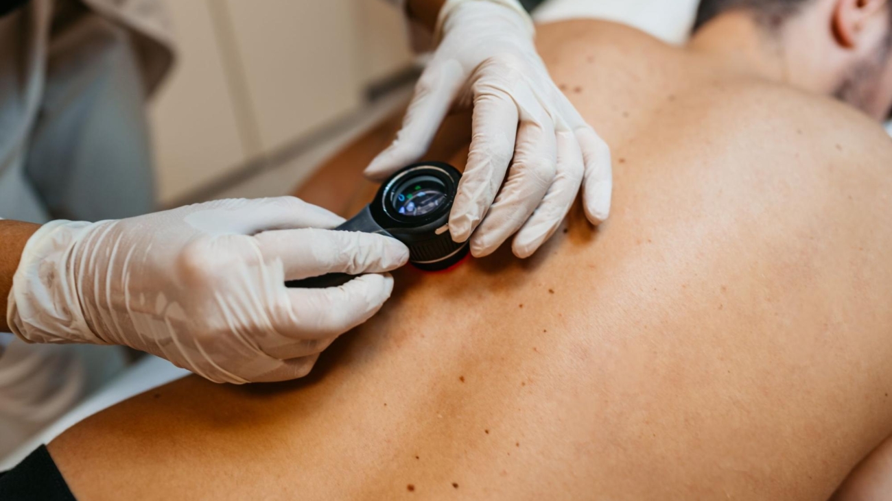
Visual inspection is the first step, but dermatologists utilize specialized diagnostic tools to examine the skin more closely. The primary instrument used in modern dermatology is the dermatoscope. This handheld device combines high-quality magnification with polarized lighting. A dermatoscope provides a magnified view of the skin surface, so the doctor can see structures invisible to the naked eye. This helps the dermatologist distinguish between harmless lesions and those requiring further investigation.
If a dermatologist identifies a suspicious lesion, they will perform a skin biopsy. During this routine procedure, the doctor removes a small sample of the affected skin tissue, and they send the sample to a laboratory, where a specialist examines it under a microscope. This microscopic analysis is the standard method for confirming the presence or absence of cancerous cells.
Call a Dermatology Practice Today
Monitoring your skin at home is helpful, but professional evaluations provide a more comprehensive approach to skin health. If you notice a new growth, a sore that fails to heal, or a mole that changes appearance, schedule a professional screening. Early identification expands the available management options and generally requires less invasive procedures. Take a proactive step toward maintaining your health; call a dermatology practice today to schedule a comprehensive skin examination.
LEAVE A COMMENT